Precision Spinal Implants, Orthopedic Power Systems & Instruments
Engineered to international regulatory benchmarks, supporting tertiary orthopedic clinics, distributors, and OEM medical brand holders across 60+ countries.
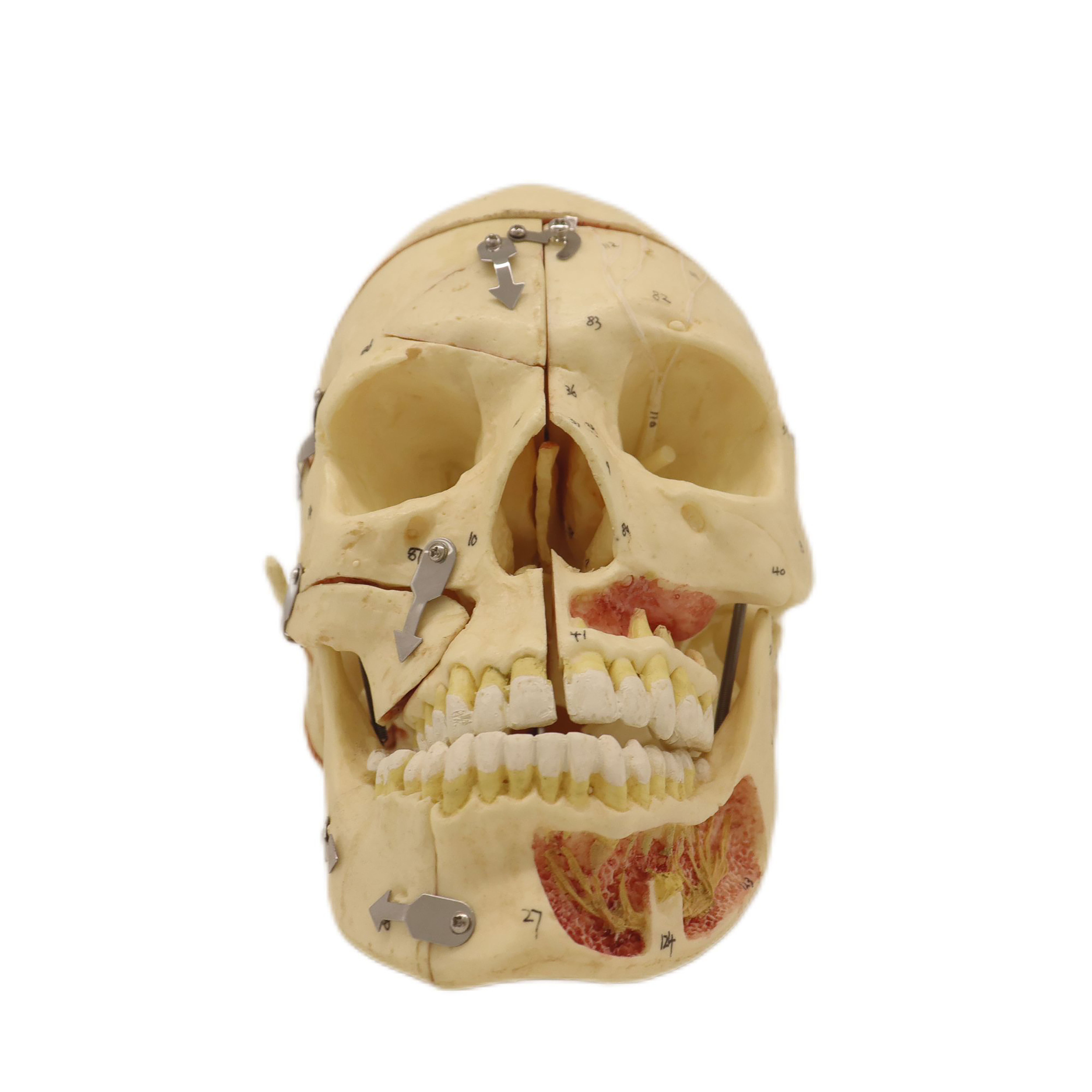
3-Part Human Muscular Skull Skeleton Model
Precision anatomical teaching specimen featuring hand-painted muscle origins and insertions, dissectible calvarium, and spring-articulated mandible for clinical training.
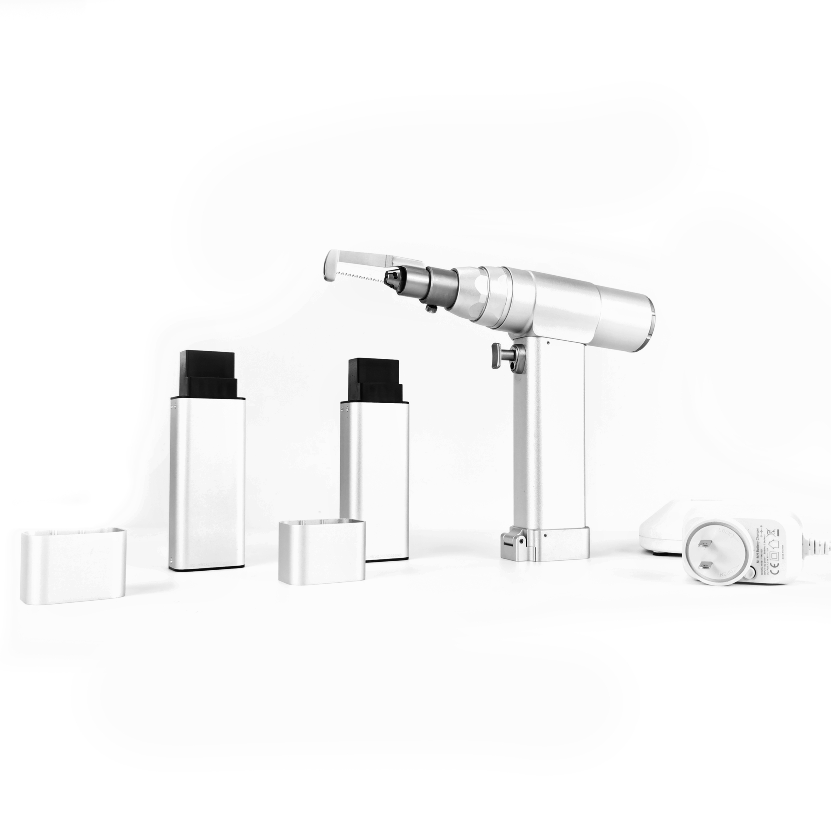
Medical Electric Sternum Saw & Orthopedic Drill
CE-compliant autoclavable motorized surgical tool system engineered with precision brushless motor, offering controlled blade oscillations and high torque for sternotomy and trauma.
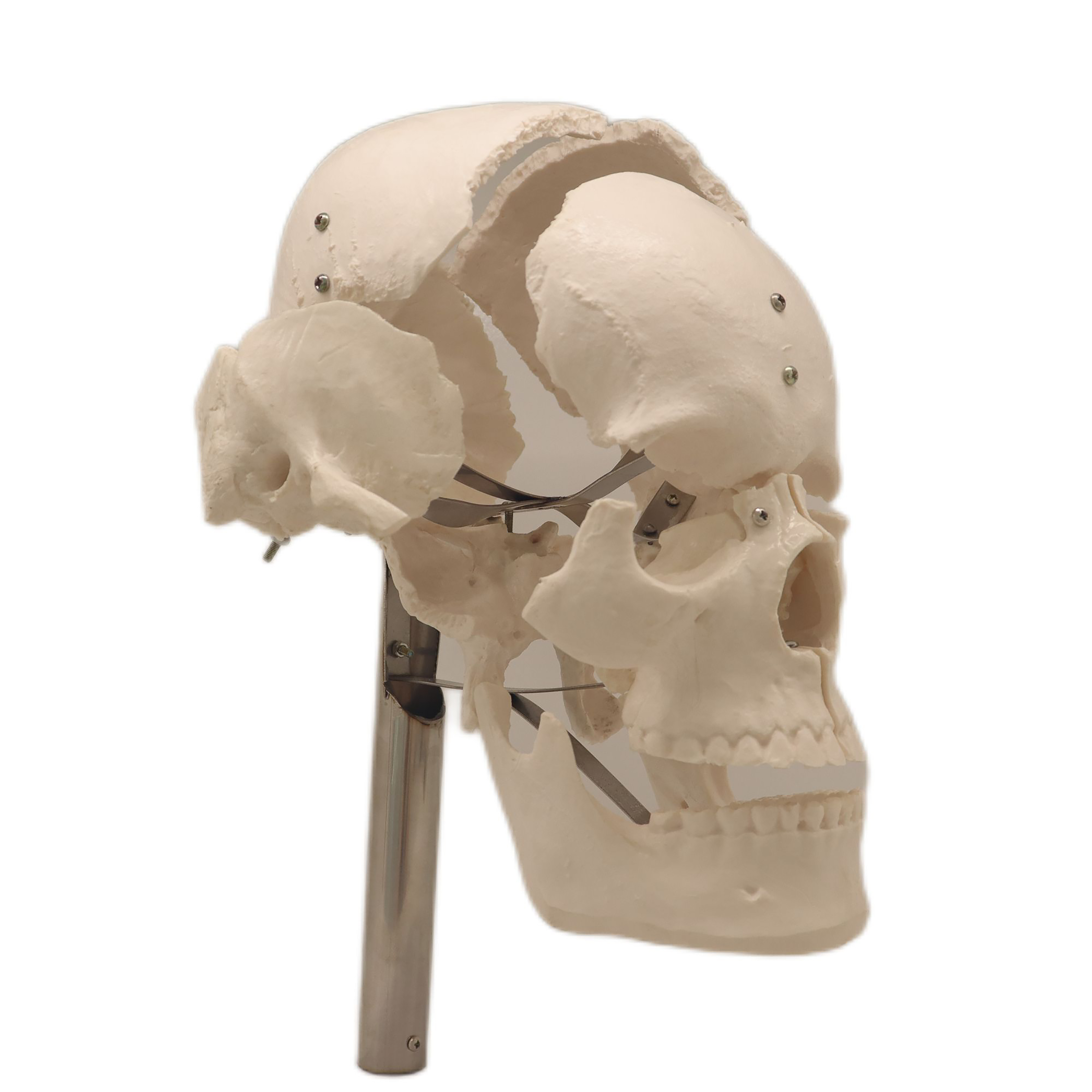
Disarticulated Cranial Bone Teaching Model
High-grade medical PVC separated skull bones designed for university anatomy labs and surgical simulation, demonstrating intricate cranial sutures, foramina, and endocranial contours.
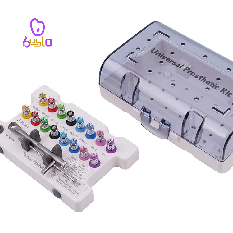
Surgical Handpiece & Implant Tool Container
Heavy-gauge stainless steel organization cassette designed for automated washing, steam autoclave protocols, and high-security containment of surgical micro-handpieces.
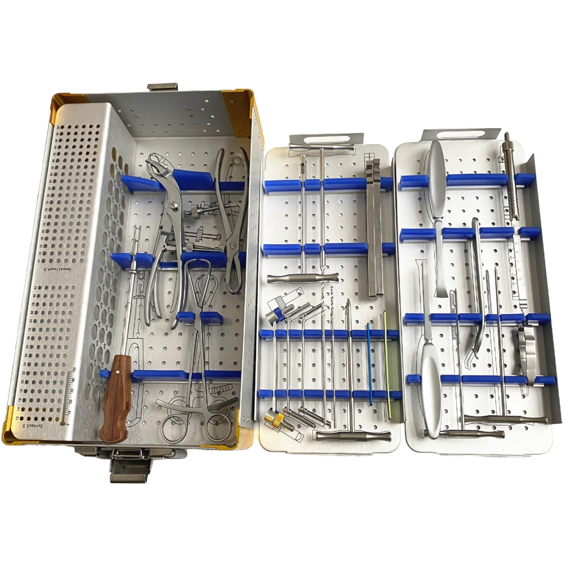
Small Fragment Upper Limb Instrument System
Turnkey surgical set incorporating high-tensile drill guides, depth gauges, reduction clamps, and tap handles optimized for radial, ulnar, and humerus plating interventions.

Ringent Rod-to-Pin Fixator Clamp Coupling
Multi-pin clamping connector crafted with medical aircraft alloy for damage-control orthopedic frames, facilitating rigid axial and rotational stabilization of open fractures.
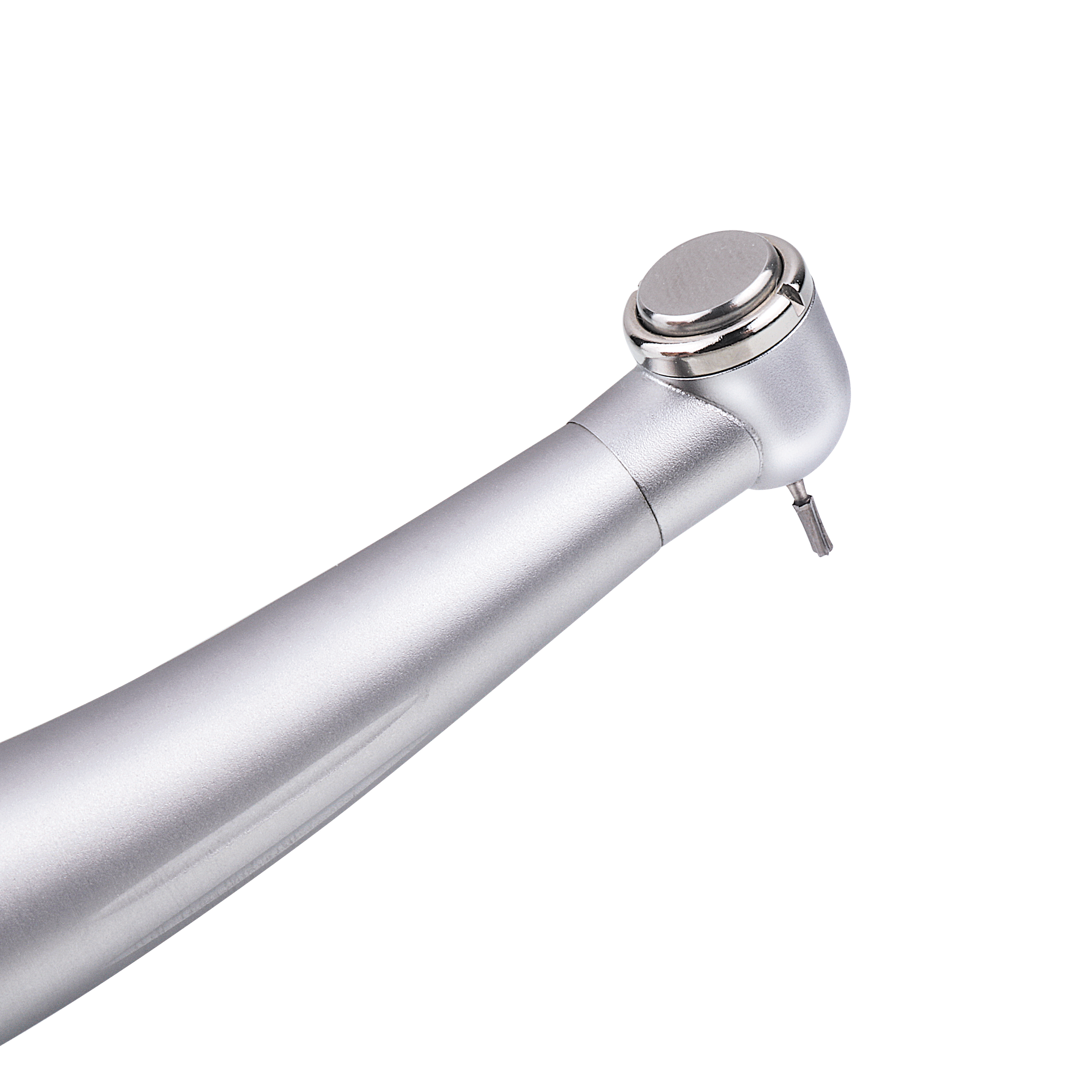
High-Speed Surgical Implant Handpiece Turbine
Precision pneumatic surgical handpiece built with ceramic bearings, dynamic balancing, and internal water spray systems, delivering zero-chatter osteotomy and bone burring.
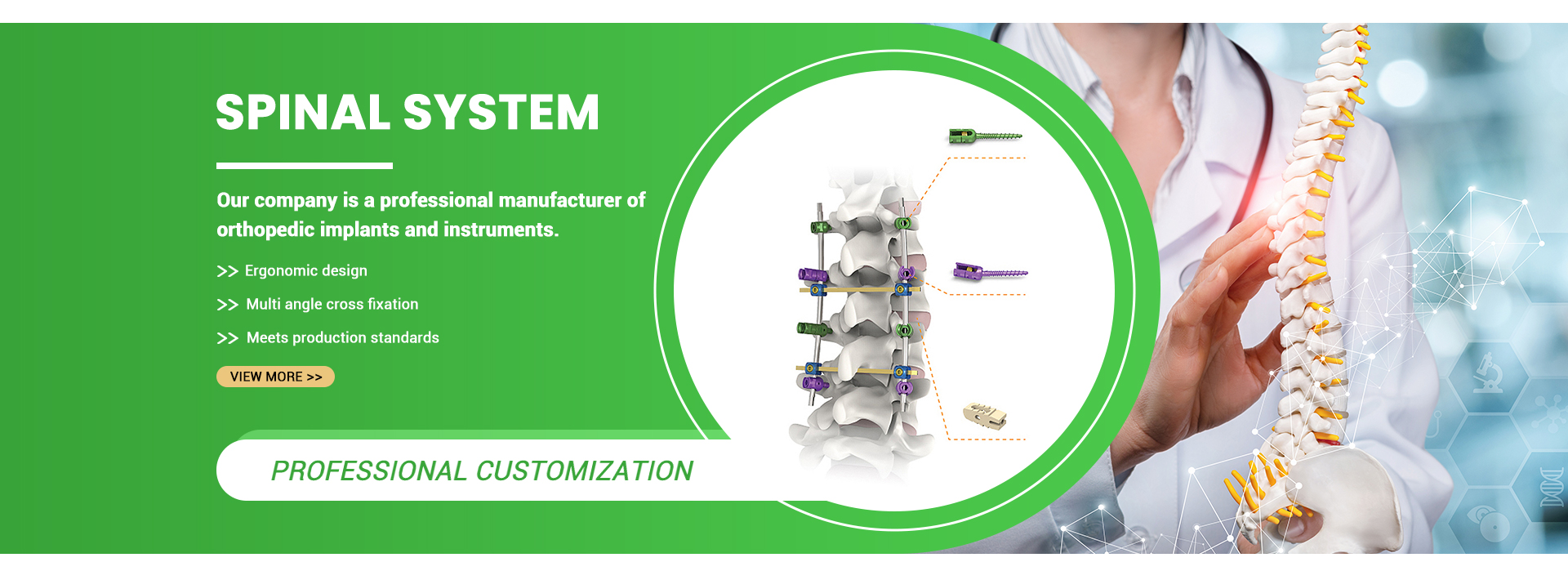
Titanium Expandable Vertebral Body Replacement
Continuous in-situ gear-expanded titanium corpectomy cage featuring modular lordotic/kyphotic endplates (0°–15°) and large graft volume for severe trauma and tumor reconstruction.
Integrated Orthopedic Production Infrastructure
From metallurgical verification in Suzhou to automated 5-axis CNC machining, Jiangsu GMV Medical Device Co., Ltd. provides clinical reliability backed by empirical testing and audited processes.
TÜV Rheinland Certified Manufacturer
Our Jinfeng Town production complex in Suzhou undergoes strict third-party inspections by TÜV Rheinland. We operate authenticated ISO 13485 quality management systems covering the full traceability lifecycle from raw ingots to sterile medical devices.
In-House 5-Axis Precision Machining
Equipped with high-precision CNC Swiss lathes, 5-axis vertical machining centers, and wire EDM slicing systems, our shop floor creates micron-level gear mechanics, anti-migration endplate spikes, and dynamic locking interfaces without outsourcing.
Complete Regulatory Dossier Support
We supply international distributors with full technical documentation: raw material melt certificates (Ti-6Al-4V ELI conforming to ASTM F136 / ISO 5832-3), biocompatibility studies (ISO 10993), static/dynamic biomechanical fatigue reports, and sterilization validations.
Biomechanics, Structural Fixation & Metallurgy of Vertebral Reconstruction
A comprehensive clinical evaluation for spine surgeons, hospital procurement committees, and orthopedic distributors qualifying medical device manufacturers in China.
1. Clinical Indications & Biomechanical Objectives of Corpectomy Reconstruction
Vertebral body replacement (VBR) devices represent the pinnacle of structural spine engineering. When catastrophic structural compromise strikes the anterior or middle spinal column due to burst fractures, high-energy trauma, severe osteomyelitis, destructive primary or metastatic tumors, or multisegmental degenerative kyphosis, total or partial corpectomy becomes imperative. The primary clinical imperative of the implant is immediate restoration of the anterior load-bearing column, preservation of sagittal alignment, prevention of late kyphotic collapse, and protection of neurologic structures from recurrent impingement.
Biomechanical load-sharing follows Wolff's Law: an ideal VBR cage must be sufficiently rigid to withstand continuous axial, torsional, and flexural loads without deformation, yet display an elastic modulus compatible with adjacent trabecular vertebral bone. Excessive structural rigidity promotes stress-shielding, impairing local biological bone incorporation and leading to implant loosening. Conversely, inadequate mechanical strength or insufficient endplate contact area precipitates catastrophic subsidence, where the implant sinks into the cancellous centrum of the adjacent vertebrae, causing kyphosis, nerve root compromise, and hardware fracture.
2. Metallurgical & Biomaterial Properties Comparison in VBR Construction
Material selection directly dictates the clinical outcome of anterior spinal column arthrodesis. At Jiangsu GMV Medical Device Co., Ltd., our engineering departments evaluate tensile strength, fatigue endurance limits under cyclic loading, elastic modulus, and radiopacity across three core implant substrate options:
| Implant Material Substrate | Elastic Modulus (GPa) | Yield Strength (MPa) | Biocompatibility & Osseointegration | Radiological Characteristics | Clinical Application Profile |
|---|---|---|---|---|---|
| Ti-6Al-4V ELI (Grade 23) ASTM F136 / ISO 5832-3 |
110 – 114 GPa | ≥ 825 MPa | Excellent biocompatibility; native TiO2 passivation layer enables robust cellular attachment. | Moderate radiopacity; slight streak artifacts on CT/MRI, manageable with artifact reduction. | Standard of care for expandable mechanical chassis, gears, locking screws, and trauma mesh. |
| Porous 3D Titanium Lattice EBM / SLM Additive Metal |
2.8 – 5.5 GPa (Matches Cancellous Bone) |
180 – 320 MPa | Superior osseointegration; 60–75% open interconnected porosity mimics natural trabecular architecture. | Permits through-growth visualization on cross-sectional MRI and high-contrast CT scans. | High-end static endplate caps, biological integration interfaces, tumor reconstruction. |
| Medical-Grade PEEK ASTM F2026 Polymer |
3.2 – 4.0 GPa | 95 – 120 MPa | Chemically inert; requires titanium plasma spray or HA coating to optimize cellular adhesion. | Radiolucent; clear assessment of internal bony bridging; requires tantalum markers. | Static cervical/lumbar spacers, multisegment tumor resections where radiation oncology is required. |
| Human Cortical Bone Reference Natural Biological Control |
12 – 18 GPa | 100 – 150 MPa | Native biological scaffold undergoing continuous vascularization and Remodeling. | Isodense with natural skeletal framework on plain radiography and computed tomography. | Autograft/Allograft fibular strut benchmark (prone to early resorption and donor morbidity). |
3. Kinematic Architecture: Expandable VBR vs. Static Titanium Mesh Cages
Corpectomy reconstruction hinges on two primary structural methodologies: static cut-to-length cylindrical mesh cages and in-situ continuously expandable mechanical cages.
Static Mesh Systems: Traditional titanium mesh cages provide substantial compressive strength and generous internal graft volume. However, they demand exact intraoperative manual trimming. An oversized mesh risks over-distraction and endplate fracture, whereas an undersized construct induces immediate instability. Furthermore, forceful impaction into the prepared disc space frequently damages the dense cortical rim of the vertebral endplates, accelerating long-term subsidence.
Continuously Expandable VBR Systems: Modern surgical protocols increasingly mandate expandable cages. These implants are introduced into the corpectomy defect at their minimal collapsed profile, maximizing clearance and minimizing the risk of iatrogenic spinal cord or nerve root laceration. Once positioned under fluoroscopic guidance, an internal rack-and-pinion, bevel-gear, or concentric screw drive is actuated via ergonomic external instrumentation. This applies controlled, measurable distraction across the defect, achieving anatomical lordosis/kyphosis correction and precise tensioning of the posterior longitudinal ligament. A secondary set-screw mechanism positively locks the expansion carriage, eliminating micro-motion or height loss during dynamic physiological fatigue cycles.
4. Endplate Contact Mechanics & Subsidence Prevention
The interface between the metallic endplate footprint and the subchondral bone of the adjacent vertebral centrum represents the primary structural failure point in spinal column reconstruction. Clinical studies demonstrate that the peripheral epiphyseal ring of the vertebra possesses significantly higher bone mineral density and ultimate compressive yield strength than the central trabecular zone.
Our engineered VBR systems incorporate wide, anatomically contoured footprint footprints (ranging from 20x24mm up to 28x34mm for thoracolumbar applications) designed to span across the peripheral cortical rim. Furthermore, directional anchoring spikes (0.8mm–1.5mm in height) achieve rigid primary mechanical stability against shearing and torsional dislodgement without compromising the integrity of the subchondral plate. Modular endplate caps offering varying lordotic and kyphotic angles (0°, 4°, 8°, 12°, 15°) ensure full surface-to-surface contact across the treated level, distributing axial loads homogeneously and eliminating localized peak stresses that initiate micro-fractures.
Future Sourcing & Technological Trends in Spinal Reconstruction
Anticipating clinical, regulatory, and metallurgical shifts shaping global spinal device tenders and distribution networks over the next decade.
1. 3D Additive Porous Architecture
The global spinal surgery sector is shifting from solid machined titanium bodies toward additive 3D electron beam melting (EBM) and direct metal laser sintering (DMLS). These techniques produce bionic, interconnected trabecular lattices with pore sizes calibrated between 500 and 700 microns. This geometry promotes rapid capillary ingrowth, osteoblast adhesion, and direct bone interlock throughout the implant core, drastically shortening fusion duration.
2. MIS & Lateral Transpsoas Approaches
Open anterior transthoracic and retroperitoneal approaches carry high morbidity. Procurement patterns highlight surging demand for minimally invasive surgery (MIS) vertebral body replacement implants compatible with lateral lumbar interbody fusion (LLIF/XLIF) corridors. Implants feature ultra-slender insertion sleeves, multi-axis radiolucent retractors, and rotating expanding drives designed to navigate through extreme micro-incisions.
3. Tactile Torque & Intelligent Navigation
Next-generation expandable corpectomy cages are being paired with intraoperative sensory instrumentation. Calibrated torque-limiting insertion wrenches provide tactile feedback to the surgeon, directly correlating turning resistance with endplate contact pressure to prevent accidental endplate penetration. Furthermore, integrated robotic optical navigation arrays allow real-time sub-millimeter positional tracking under 3D intraoperative CT fluoroscopy.
Global Supply Chain Restructuring & Direct Factory Qualification
International healthcare procurement organizations, hospital consortia, and private label medical device enterprises are systematically restructuring their orthopedic vendor ecosystems. In response to inflationary pressures, escalating European MDR compliance overheads, and supply-chain vulnerabilities, global buyers are pivoting away from multi-tiered distributor intermediaries to forge direct, long-term partnerships with verified Tier-1 Chinese manufacturers.
Qualifying an overseas spinal implant partner demands rigorous scrutiny. Established institutions prioritize manufacturers offering integrated in-house metallurgical production, raw-material lot traceability, automated cleaning validated under ISO 13408 cleanroom standards, and transparent third-party facility audits. By establishing direct OEM/ODM supply agreements with Jiangsu GMV Medical Device Co., Ltd., procurement teams achieve significant cost efficiencies, direct access to senior design engineers, expedited prototyping cycles, and protected supply line continuity.
Frequently Asked Questions: Sourcing Spinal Implants from China
Detailed technical, regulatory, and logistical responses to common queries from global procurement officers, distributors, and orthopedic hospital networks.
What regulatory certifications and testing dossiers accompany GMV vertebral body replacement systems?
How does GMV ensure metallurgical purity and raw material traceability?
What OEM and ODM customization capabilities does your facility support for spinal implants?
Are dedicated surgical instrument sets supplied with the vertebral body replacement implants?
What are the minimum order quantities (MOQ), production lead times, and export logistics?
Can international hospital procurement committees audit your manufacturing plant?
How do GMV expandable cages address endplate subsidence and intraoperative dislodgement?
Request Complete Product Catalogs, CAD Models & Distributor Pricing
Partner directly with Jiangsu GMV Medical Device Co., Ltd. for spinal internal fixation, expandable vertebral body replacement systems, and customized OEM orthopedic instrumentation. Connect with our engineering and export team today.
